Pale spots after pico laser for melasma
Hypopigmentation Following Picosecond Laser Treatment for Melasma: A Case Series
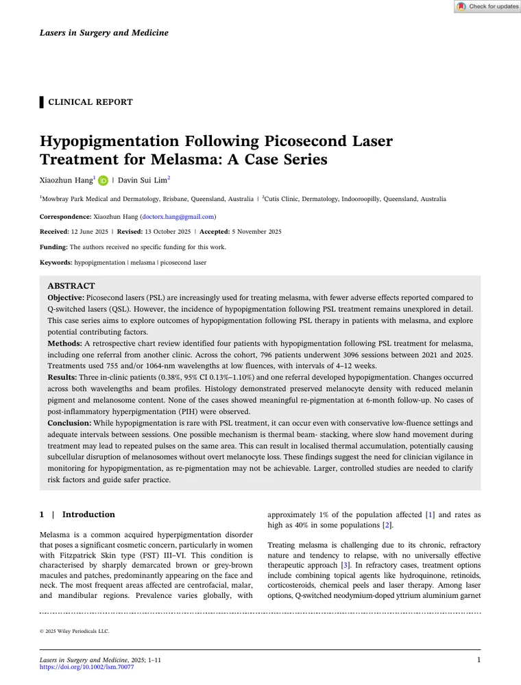
Four patients treated with picosecond laser for melasma developed pale spots that had not recovered six months later. The biopsies showed the pigment cells were still alive — they had simply stopped making pigment.
Four numbers worth knowing
What we found
It is rare, but it is real.
Across 796 patients and 3,096 treatment sessions between 2021 and 2025, three in-clinic patients developed hypopigmentation, with one further case referred from another clinic. That is 0.38%, with a confidence interval from 0.13% to 1.10%.
None of it recovered.
At six months, not one of the four showed meaningful repigmentation. No cases of post-inflammatory hyperpigmentation — the complication everyone watches for — were observed at all.
The pigment cells survived.
Biopsies showed preserved melanocyte density with normal dendritic processes, alongside reduced epidermal melanin and reduced melanosome content. The cells were present and structurally intact; what had failed was pigment production and delivery.
It happened at conservative settings.
Fluences ranged from 0.4 to 1.0 joules per square centimetre with intervals of four to twelve weeks — settings within the range generally regarded as safe. These were not aggressive treatments.
Pulse stacking is the likely mechanism.
Slow hand movement during treatment leads to repeated pulses landing on the same area. That causes localised thermal accumulation, which can disrupt melanosomes without killing the melanocyte — exactly what the histology showed.
It occurred across wavelengths and beam profiles.
Both 755 nm and 1064 nm were involved, and both flat optic and fractional beam modes. The problem does not appear to belong to one setting on one machine.
What this means for you
Ask about hand speed and overlap, not just the machine
The implicated factor here is technique rather than device. Repeated passes over the same spot within a session, and slow movement across the skin, are what drive thermal accumulation.
Conservative settings are not a guarantee
These cases occurred at low fluences with adequate intervals between sessions. Low-and-slow is safer than aggressive, but it is not risk-free, and it should not be described as though it were.
This one may not reverse
None of the four repigmented at six months. When you consent to pico laser for melasma, treat the possibility of a permanent pale patch as real, even though it is uncommon.
Photographs between sessions
All four cases were confirmed on serial standardised photography. It is how you catch focal lightening while it is still small enough to change course.
Why this matters
Picosecond lasers have become the preferred device for melasma in much of the world, and in Asia low-fluence treatment is common enough to have its own name. The appeal is real: shorter pulses, less heat spreading into surrounding tissue, and far less of the post-inflammatory darkening that followed Q-switched treatment.
Hypopigmentation after Q-switched lasers is a well-recognised side effect, reported in up to 21% of published studies, and it can be permanent and disfiguring. The assumption has been that picosecond technology is largely free of it. This is the first case series to document it happening for melasma specifically.
The distinction that matters
Melanocyte survival is not the same as pigment safety. A cell can be present, structurally normal and still no longer doing its job — which is why counting melanocytes underestimates the risk.
Who was studied
A retrospective chart review of every patient treated with picosecond laser for melasma at one Australian clinic between January 2021 and August 2025, identifying those who developed hypopigmentation and examining treatment parameters, histology and six-month outcomes.
- Patients treated
- 796
- Sessions
- 3,096
- Cases identified
- 4
- Wavelengths
- 755 nm and 1064 nm
- Follow-up
- 6 months
What this study cannot tell you
Every study has a boundary, and it is fairer to state it than to leave you to guess.
- Four cases. That is a case series, and it is presented as one — it describes what happened, not how often it will happen to you.
- Retrospective chart review, so treatment parameters were recorded as part of routine care rather than for study purposes.
- One of the four was referred from another clinic, and those treatment parameters were not documented.
- Follow-up stopped at six months. Whether these areas eventually repigment over years is unknown.
- Pulse stacking is a proposed mechanism consistent with the histology, not a demonstrated cause. Larger controlled studies are needed to identify the real risk factors.
Questions people actually ask
How common is hypopigmentation after picosecond laser?
In this series, 0.38% — three cases among 796 patients treated across 3,096 sessions, with a confidence interval running from 0.13% to 1.10%.
Uncommon, but not negligible, and worth knowing before a course of treatment rather than after it.
Will white spots after laser go away?
None of the four patients in this series showed meaningful repigmentation at six months.
The biopsies offer some grounds for hope, in that the pigment cells were still present and structurally normal rather than destroyed — so recovery is biologically possible. But nothing here demonstrates it, and it should be treated as potentially permanent when you are weighing up treatment.
Is low-fluence laser toning safe for melasma?
It is widely used and generally well tolerated, with notably less post-inflammatory darkening than Q-switched treatment — no cases of that occurred in this entire series.
The caution is that these hypopigmentation cases happened at conservative fluences with adequate intervals. Lower energy reduces risk; it does not remove it, and the cumulative exposure across many sessions appears to matter as much as the setting on any single one.
Cite this paper
Hang X, Lim DS. Hypopigmentation Following Picosecond Laser Treatment for Melasma: A Case Series. Lasers in Surgery and Medicine. 2025. doi:10.1002/lsm.70077
Related reading
Want this assessed properly?
A virtual consultation gives you a written report and a plan before we speak.
Published 2025 · Summary written and reviewed by Dr Davin Lim, FACD · General information, not personal medical advice.
Have some questions?
Send a message and it will be answered personally. Form endpoint — to be wired in production